Figure 5.

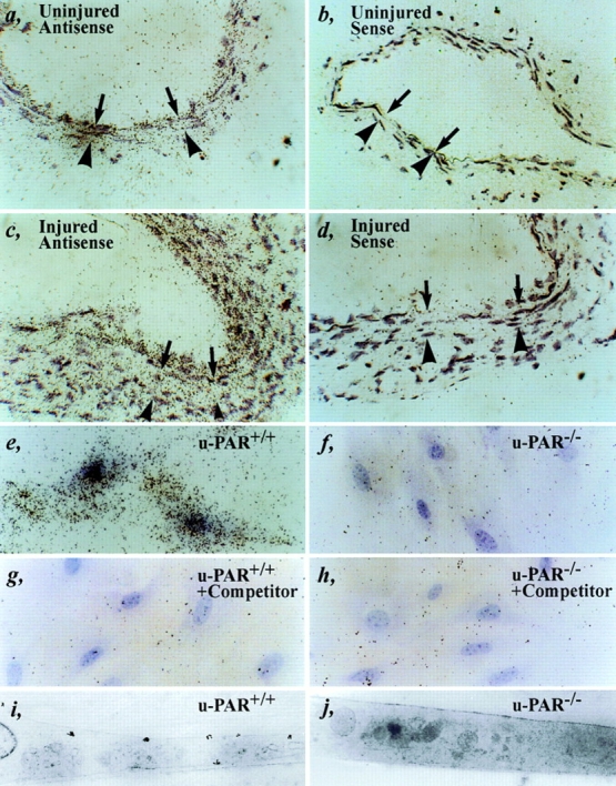
Expression of u-PAR. (a–d) In situ hybridization of an uninjured (a and b) or injured (c and d) wild-type artery within 1 wk after injury (at locations 1–3 or 8–10 as defined in Fig. 1) with an antisense (a and c) or sense (b and d) murine u-PAR probe, revealing a low basal level of u-PAR mRNA expression in a quiescent artery and significantly induced u-PAR mRNA levels at the leading front of cellular migration. The arrows indicate the internal elastic lamina; the arrowheads indicate the external elastic lamina. (e–j) Autoradiography (light microscopy: e–h, or electron microscopy: i and j) of cultured u-PAR+/+ (e, g, and i) and u-PAR−/− (f, h, and j) smooth muscle cells, incubated with 2 nM murine 1–48u-PA alone (e, f, i, and j) or together with a 100-fold excess of unlabeled competitor (g and h), revealing significant binding to u-PAR+/+ but not to u-PAR−/− smooth muscle cells. Bar, 25 μm.
