
Figure 6.
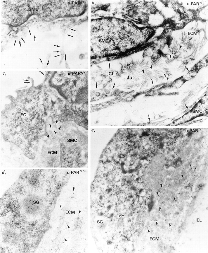
Ultrastructural localization of u-PA in vivo. Immunogold labeling of u-PA in u-PAR+/+ (a–c) and u-PAR−/− (d and e) arteries, revealing u-PA bound to the cell surface (a–c, arrows) on endothelial cells (EC) or smooth muscle cells (SMC), or associated with the extracellular matrix (ECM) (b and c, arrowheads) in u-PAR+/+ arteries. Note the presence of u-PA gold particles on cell projections (a), and on collagen (CL) fibers in the extracellular matrix (b). In contrast, in u-PAR−/− arteries, u-PA gold particles were predominantly detected in the pericellular milieu associated with extracellular matrix components (d and e, arrowheads). Note the paucity of u-PA gold particles on the elastin fibers of the internal elastic lamina (IEL) (e). Gold particles labeling u-PA were also found in secretory granules inside u-PAR−/− cells (d and e, SG).