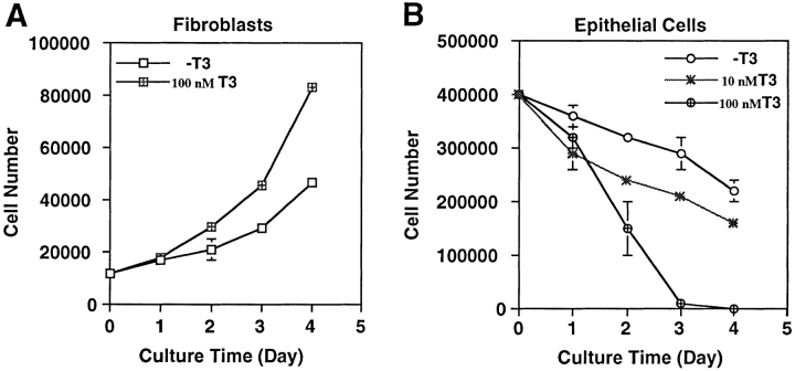
Figure 1.
Contrasting effects of thyroid hormone on tadpole intestinal epithelial and fibroblastic cells. The fibroblasts (A) and epithelial cells (B) were isolated from stage 57/58 of tadpole small intestine and then cultured on a six-well plastic dish in 60% L-15 medium containing 10% T3- depleted fetal bovine serum at 25°C in the presence or absence of 10 or 100 nM T3. The live cells were counted daily by trypan blue staining. Note that the epithelial cell number decreased even when no exogenous T3 was present. This could be because of the residual T3 in the treated serum. DNA fragmentation and flow cytometry analyses (Figs. 2 and 3) indicated that at least part of this decrease was due to apoptosis.

