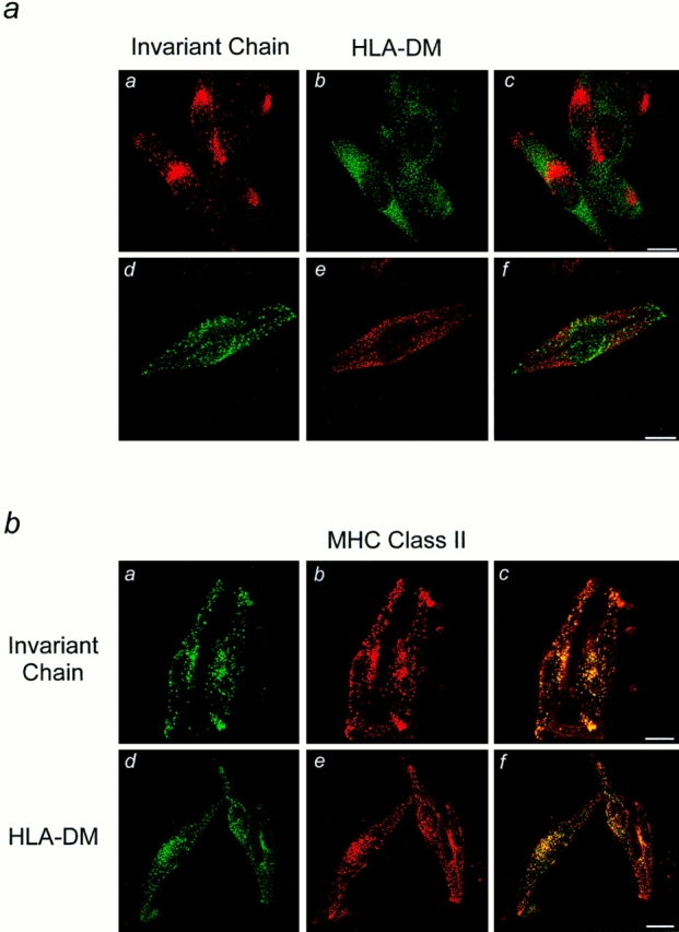
Figure 4.

Localization of Ii and HLA-DM by immunofluorescence microscopy. (a) Cells grown on coverslips were fixed, permeabilized and labeled for Ii using anti-Ii NH2-terminal monoclonal (a) or polyclonal (d) antibodies, and HLA-DM using polyclonal (b) or monoclonal antibodies (e) followed by FITC (b and d) or Texas red (a and e) conjugated secondary antibodies. The superposition of the images is shown in c and f. In b, cells were labeled for MHC class II molecules using monoclonal (b) or polyclonal (e) antibodies and double labeled for Ii using polyclonal anti-Ii NH2-terminal antibodies (a) or HLA-DM using mAbs (d), followed by Texas red (b and e) or FITC (a and d) conjugated secondary antibodies. c and f show the merged images. Bars: 20 μm.
