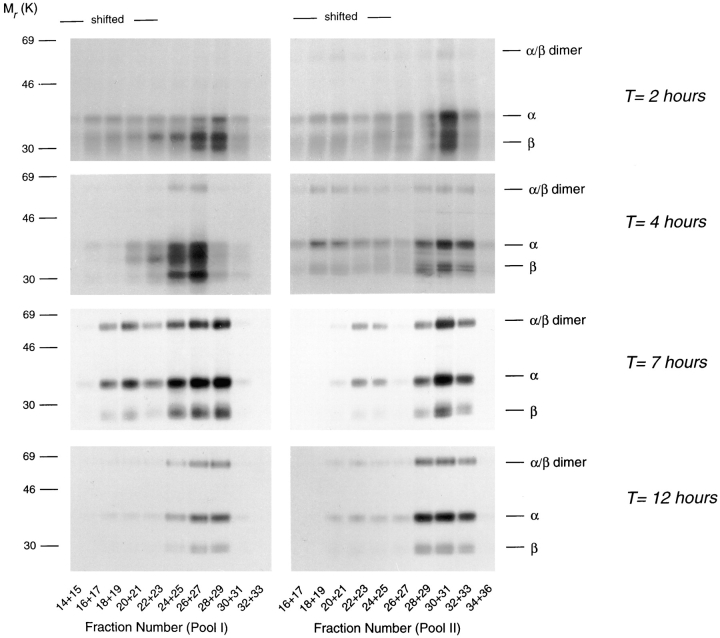
Figure 6.
Formation of SDS-stable αβ dimers in class II–containing subcellular organelles. Cells were labeled for 20 min with [35S]methionine/cysteine, followed by a chase for the times indicated in the absence of radiolabel. At each time point, cells were homogenized and membranes fractionated by sucrose density centrifugation and organelle electrophoresis as described in Fig. 2. Fractions after organelle electrophoresis were pooled as indicated, and class II molecules immunoprecipitated followed by SDS-PAGE under mildly denaturing conditions and fluorography. The protein peak migrated at fraction 26 (Pool I) and fraction 30 (Pool II), respectively. Note that the fluorographs represent equal amount of the class II signal in pool I and II (see text).

