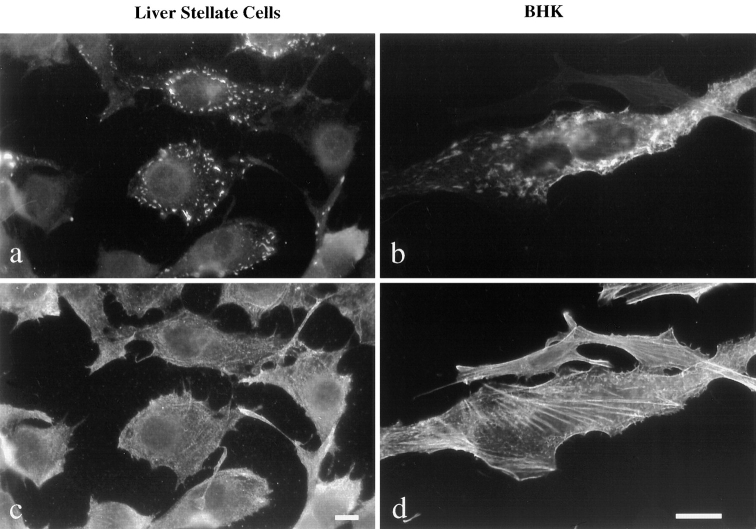
Figure 2.
Localization of nonmuscle and sarcomeric MyHC expressed in liver stellate cells and BHK cells. Indirect immunofluorescence was performed to visualize MyHC in liver stellate cells (a and c) and BHK cells (b and d). Sarcomeric MyHC (a and b) forms punctate structures around the plasma membrane of liver stellate cells whereas it is more uniformly distributed in the cytoplasm of BHK cells. Sarcomeric (a and b) and nonmuscle MyHC (c and d) show partial colocalization in BHK cells, but not in liver stellate cells. Bars, (a and c) 10 μm, and (b and d) 20 μm.

