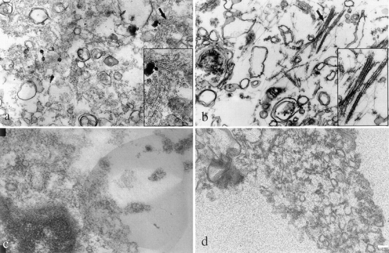
Figure 4.

Ultrastructural analysis of sarcomeric MyHC in liver stellate cells and BHK cells. Immunoelectron microscopy was performed to determine the organization of sarcomeric MyHC in liver stellate cells and BHK cells. Monoclonal antibody, F59, was used to detect sarcomeric MyHC in liver stellate (a) and BHK cells (b); anti–α-platelet myosin antibody was used to detect nonmuscle MyHC in liver stellate cells (c) and BHK cells (d). Secondary antibodies were 5 nm gold-conjugated IgG. Arrows denote areas in inset for a and b. Bar, 1 μm.
