
Figure 7.
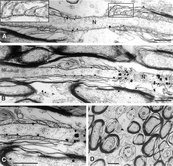
Immunoelectron microscopic localization of Caspr in myelinated and unmyelinated nerve fibers of the corpus callosum. Representative longitudinal sections of nodal regions of myelinated fibers with silver enhanced immunogold particles denoting the distribution of Caspr are shown in A–C. Gold particles were concentrated along the inner surface of the axonal membrane beneath the septate-like junctions located in the paranodal region (A–C). The inset in A shows the septate-like junctions that form between the axon and paranodal glial loops at higher magnification. Extremely dense labeling was observed where the plane of section approached the inner surface of the axonal membrane at the paranodes (pn; B and at higher magnification of the same field in C). Gold particles were occasionally found associated with the axonal membrane or cytoplasm in internodal portions of the myelinated fibers (B and C) but were rarely seen in the node (N). (D) A cross section demonstrating labeling of small caliber axons that are unmyelinated (u). Bars, 0.5 μm.