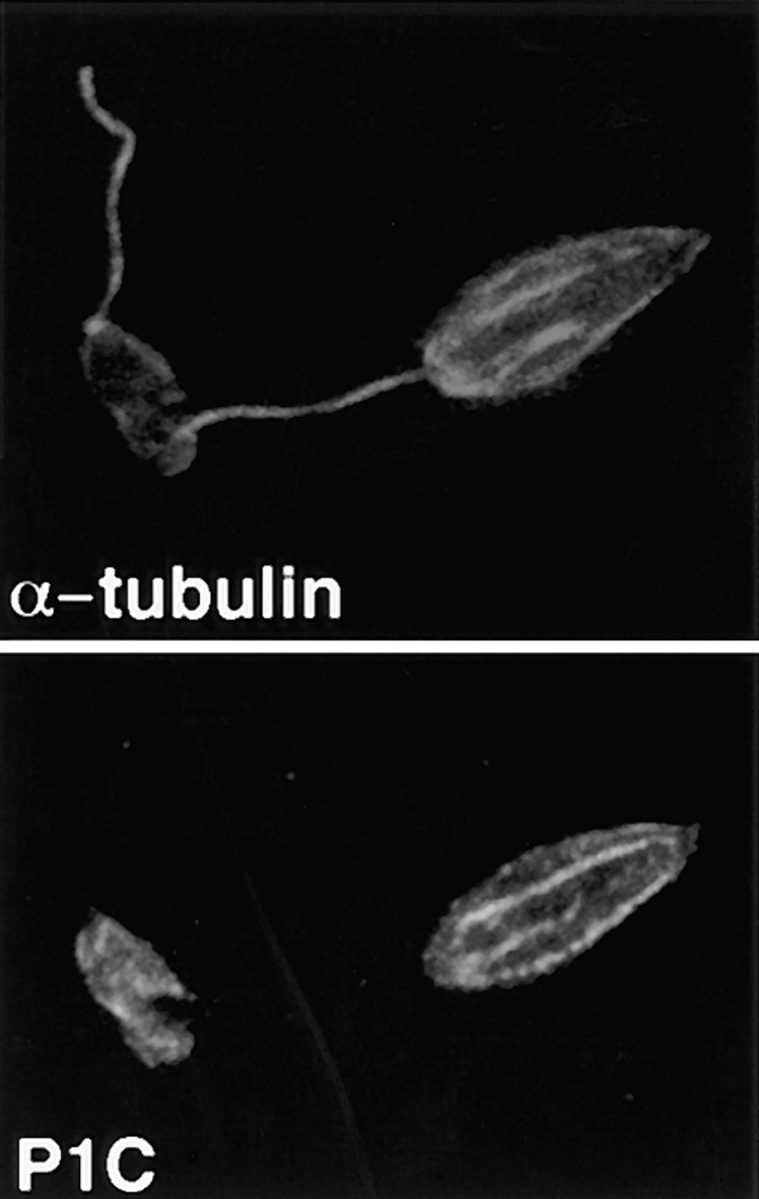
Figure 3.

Double-label confocal laser scanning micrographs of Triton X-100–extracted L. enriettii promastigotes stained with anti-P1C and anti– α-tubulin. Cytoskeletons were fixed with methanol, stained with a 1:100 dilution of the anti-P1C antibody and a 1:500 dilution of the anti–α-tubulin antibody, and then with an FITC-conjugated anti–rabbit IgG (P1C) and a rhodamine-conjugated anti–mouse IgG (α-tubulin) secondary antibody. Cytoskeletons were examined by confocal microscopy using illumination at 488 nm to visualize the Pro-1 glucose transporter–complexed FITC antibody (P1C) or at 546 nm to visualize α-tubulin complexed with the rhodamine antibody (α-tubulin). Each micrograph represents a single 0.5-μm section through each field.
