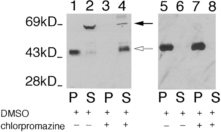
Figure 4.
Immunoblot of pellet (P) and the supernatant (S) fractions from L. enriettii incubated with 10% DMSO (lanes 1, 2, 5, and 6) or 1 mM chlorpromazine in 10% DMSO (lanes 3, 4, 7, and 8), followed by Triton X-100 extraction. The samples were run on an 8% acrylamide gel. The blot containing lanes 1–4 was probed with the anti-P1C antibody (1:100), and the blot containing lanes 5–8 was probed with anti–α-tubulin antibody (1:1,000). The solid arrow indicates the position of iso-1, and the open arrow indicates the position of iso-2. The numbers at the left indicate the migration of molecular mass markers, with molecular masses given in kD.

