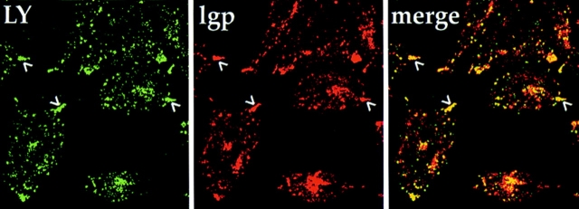
Figure 9.
Peripheral lysosomes are accessible to fluid-phase markers in ldlF cells. To determine if the peripheral lysosomes in Fig. 7 were accessible to a fluid-phase tracer, cells were first incubated for 6 h at 40°C and then labeled with LY for 90 min and washed for 90 min at 40°C. The cells were then fixed and stained for lysosomes using the lgp-B antibody shown in Fig. 7. In ldlF cells, the lysosomes (red) are found at the tips of the cells (arrowheads), but these structures are still accessible to internalized LY (green) as indicated by the yellow color in the merge panel.

