
Figure 2.
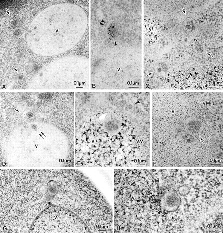
Membrane-bound forms of Cvt complex in TVY1 (pep4) cells. Cells were grown in YEPD medium at 30°C to log phase and prepared for electron microscopy as described in Fig. 1. (A and B) Immunostaining image showing Cvt vesicles (marked with arrows in A) in the cytosol. Arrowhead and double arrowheads show the inner and outer membrane of Cvt vesicle, respectively. (C) Freeze-substitution fixation image of Cvt vesicles (marked with arrows). (D) Immunostaining of Cvt vesicle (marked with arrows) in the cytosol and Cvt body (marked with double arrows) in the vacuole. (E and F) Freeze-substitution fixation image of Cvt bodies in the vacuole (marked with arrows). A Cvt vesicle is marked with an arrowhead. (G and H) Freeze-substitution fixation image of Cvt vesicle contacting and fusing to a vacuole. V, Vacuole; VM, vacuolar membrane.