
Figure 4.
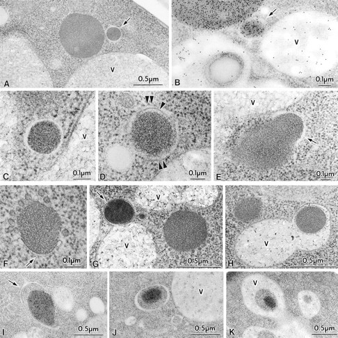
Transport of Cvt complex in SEY6210 (wild-type) harboring 2μ plasmid encoding API under growing and nitrogen starvation conditions. Vegetative cells grown in SD medium were observed (A–E). For starvation, logarithmically growing cells in SD were transferred to SD(-N) for 25 min (F–H) or 1 h (I–K) in the presence of 1 mM PMSF. Samples were prepared for electron microscopy as described in Fig. 1. (A) Freeze-substitution fixation image of Cvt vesicle (marked with an arrow). (B) Immunostaining of a Cvt vesicle (marked with an arrow). (C and D) Higher magnification image of Cvt vesicle. Arrowhead and double arrowheads show the inner and outer membrane of a Cvt vesicle, respectively. (E) Freeze-substitution fixation image of a membrane sac enclosing small portion of a large Cvt complex (marked with an arrow). (F) Freeze-substitution image of the wrapping of a Cvt complex by the isolation membrane (marked with an arrow). (G) Freeze-substitution image of a Cvt vesicle (marked with an arrow) fusing to a vacuole. (H) Freeze-substitution image of autophagic body containing a Cvt complex. (I) Immunostaining image depicting the wrapping of a Cvt complex. The arrow marks the enwrapping membrane. (J) Immunostaining of an autophagosome containing a Cvt complex. (K) Immunostaining of an autophagic body containing a Cvt complex in the vacuole. V, vacuole.