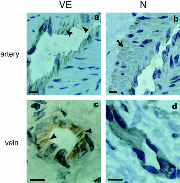
Figure 2.
Immunohistological localization of VE- (a and c) and N-cadherins (b and d) in an artery and a vein from human lymph node tissue sections. VE-cadherin is localized at cell–cell contacts (arrowheads) whereas N-cadherin staining was always diffuse in endothelial cells. Note the positive staining of arterial smooth muscle cells (arrow). Bar, 50 μm.

