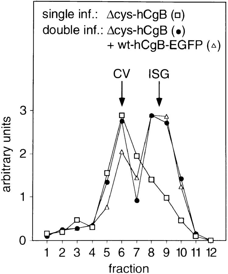
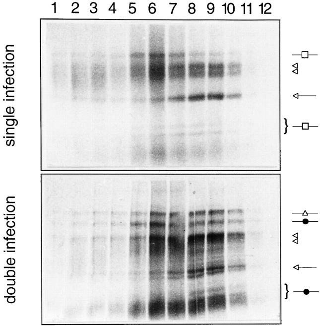
Figure 13.
Vaccinia coexpression of full-length hCgB rescues sorting of Δcys-hCgB to ISG. PC12 cells were either single- infected with vv:Δcys-hCgB (top, bottom) or double-infected with vv:Δcys-hCgB and vv:wt-hCgB-EGFP (middle, bottom), pulsed for 5 min with [35S]sulfate and chased for 12 min. Postnuclear supernatants were prepared and fractionated by sequential, velocity, and equilibrium sucrose gradient centrifugation. Equal aliquots of the equilibrium gradient (fraction 1, top) were analyzed by SDS-PAGE and phosphoimaging. Top: single infection; □, position of Δcys-hCgB and fragments (curly bracket, see also Fig. 10, middle). Middle: double infection; ▵, position of wt-hCgB-EGFP; •, position of Δcys-hCgB and fragments (curly bracket, see also Fig. 10, middle). Top, middle: endogenous hsPG and rSgII of noninfected cells are indicated by open double arrowheads and open arrows, respectively. Bottom: quantitation of virally expressed recombinant proteins across the equilibrium gradients shown in top and middle panels. The position of CV and ISG is indicated.


