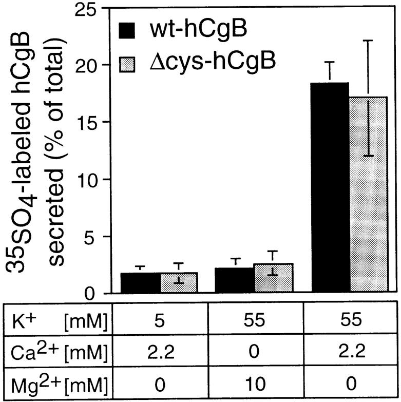
Figure 4.
Depolarization- induced, calcium-dependent secretion of wt-hgB and Δcys-hCgB. PC12 cells transfected with wt-hgB or Δcys-hCgB were pulse labeled with [35S]sulfate for 10 min and chased for 90 min. Cells were then incubated for 10 min, as indicated. Equal aliquots of cells and media were analyzed after immunoprecipitation by SDS-PAGE and phosphoimaging. The graph shows the quantitation of [35S]sulfate-labeled hCgB secreted during the 10-min incubation. Each value is expressed as percentage of total (sum of cells plus medium). Error bars show standard deviation of four independent experiments.

