Figure 5.
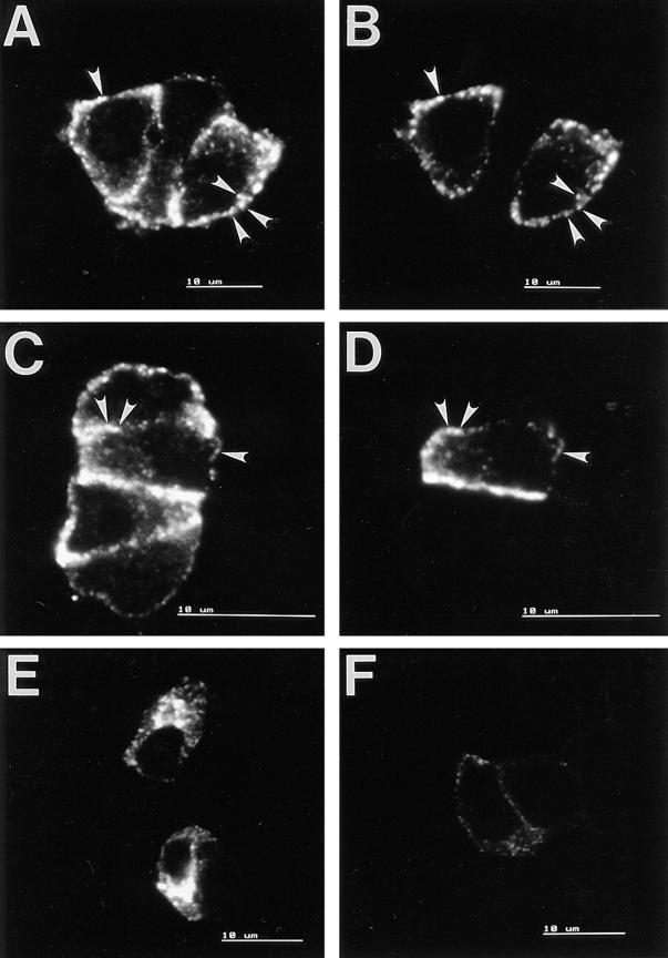
Confocal immunofluorescence analysis of PC12 cells transfected with wt-hCgB, Δcys-hCgB, and AT. Single sections are shown. A–D: PC12 cells were transfected with wt-hCgB (A and B), or Δcys-hCgB (C and D), incubated with cycloheximide for 90 min, and analyzed by double immunofluorescence for endogenous rCgB (A and C) or transfected hCgB (B and D). Note the colocalization of wt-hCgB and Δcys-hCgB with rCgB (arrowheads). E and F: PC12 cells were transfected with AT, incubated without (E) or with (F) cycloheximide for 90 min, and analyzed by single immunofluorescence for AT. Micrographs E and F were obtained with the same confocal setting and exposure time. Bars, 10 μm.

