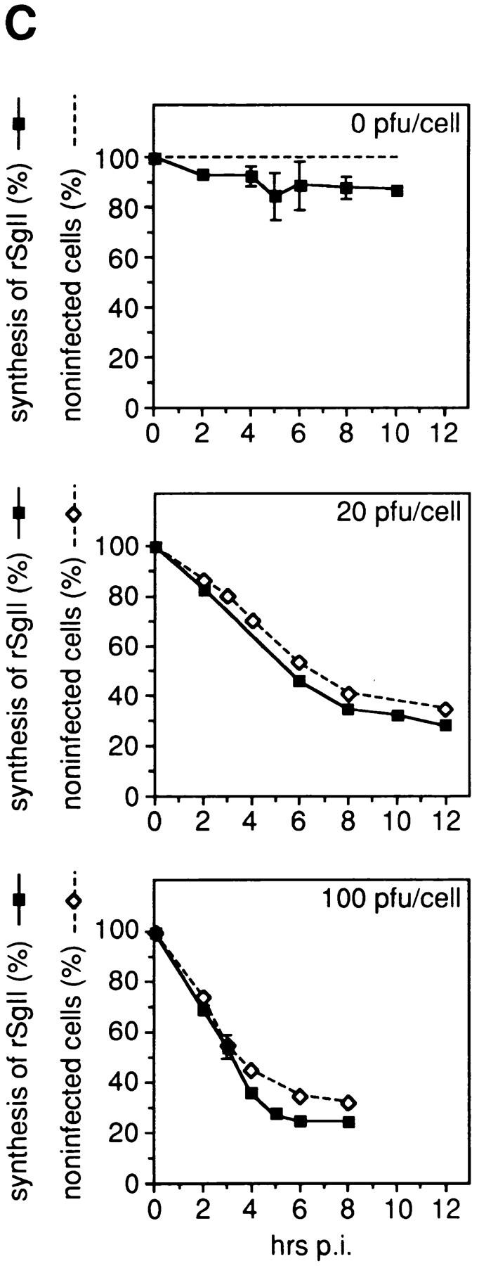
Figure 6.


Vaccinia virus infection of PC12 cells blocks endogenous protein synthesis. In A and B, PC12 cells were either mock-infected (m) or infected with vv:wt-hCgB (vv) for 8 h with 20 pfu/ cell. (A) Cells were pulse labeled for 5 min with [35S]methionine. Equal aliquots of cell homogenates (total) and of heat stable fractions prepared therefrom (HS) were analyzed by SDS-PAGE and fluorography. Full-length hCgB, arrows; rCgB, bracket; rSgII, arrowhead. (B) Immunoblot of cell homogenates (total) for hCgB (arrow). (C) PC12 cells were either mock-infected (0 pfu/cell) or infected with vv:wt-hCgB at 20 or 100 pfu/cell. After the indicated time periods of infection (hrs p. i.), cells were either fixed and analyzed for infection by immunofluorescence for hCgB, or analyzed for the synthesis of rSgII by metabolic labeling with [35S]methionine. The percentage of noninfected cells, defined by the lack of hCgB immunofluorescence (broken lines with diamonds), or the extent of rSgII synthesis, expressed as percentage of that obtained before infection (0 hrs p. i.; continuous lines, filled squares) are shown. Data are from single experiments (0 pfu, 0, 2, 10 hrs p. i.; 20 pfu, all time points; 100 pfu, 0, 6, 8 hrs p. i.), or are the mean of duplicate experiments (0 pfu/cell, 4, 5, 6, 8 hrs p. i.), or of triplicate experiments (100 pfu, 2, 3, 4 hrs p. i.). Error bars indicate SEM or the variation of individual values from the mean.
