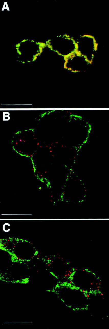
Figure 7.

Secretory granules formed under vaccinia infection lack endogenous granins. PC12 cells were infected with vv:wt-hCgB, and then chased for 90 min with cycloheximide, and, thereafter, analyzed by confocal double-immunofluorescence. Single sections are shown. A monoclonal antibody specific for virally expressed hCgB and polyclonal antibodies specific for endogenous rCgB or rSgII were used. (A) Red: α-rCgB, green: α-rSgII. (B) Red: α-hCgB, green: α-rCgB. (C) Red: α-hCgB, green: α-rSgII. Note yellow signal in A reflecting colocalization of endogenous granins and red and green signals in B and C, reflecting distinct localization of virally expressed hCgB and endogenous granins.
