
Figure 6.
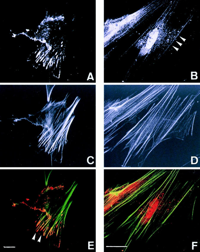
Confocal fluorescence microscopy showing actinins and the actin cytoskeleton in uterine endometrial fibroblasts. (A, C, and E) Double fluorescence of actinin-1 detected by BM-75.2 mAb (A and red in E) and actin by phalloidin-conjugated FITC (C and green in E) is shown. Actinin-1 is localized at the ends of actin stress fibers (E, arrowheads). (B, D, and F) Double fluorescence of actinin-4 detected by NCC-Lu-632 mAb (B and red in F) and actin by phalloidin-conjugated FITC (D and green in F) is shown. Actinin-4 is colocalized specifically with actin stress fibers (B, arrowheads). Bars, 5 μm.