
Figure 10.
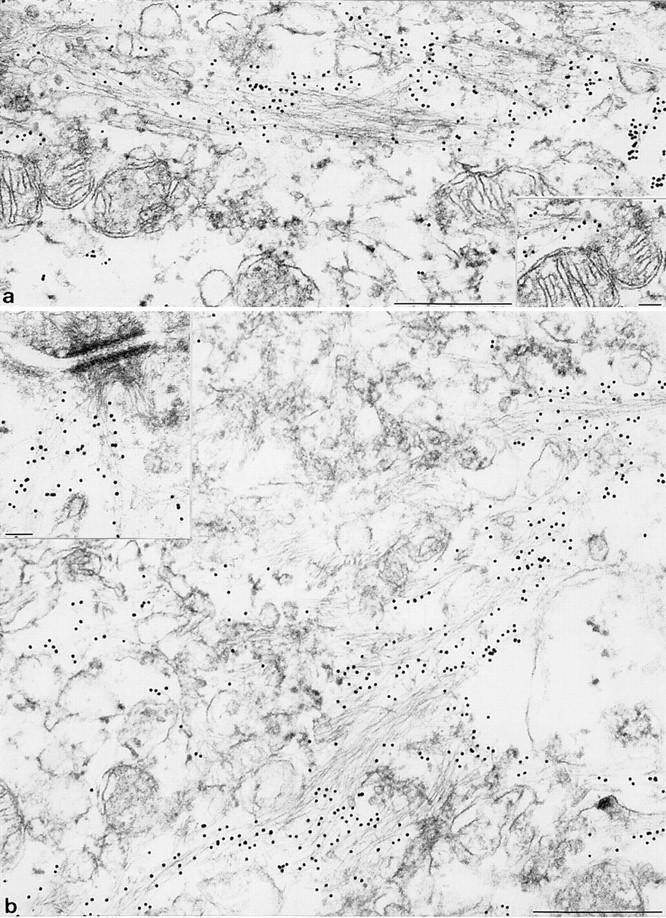
Immune electron microscopy of keratin IF in uterine epithelia from wild-type (a) and K18 null mice (b). Wild-type mice express K7, 8, 18, and 19 in uterus epithelium. (a) IF arranged in loose bundles were identified as keratins after labeling with specific primary antibodies followed by secondary antibodies coupled to 10 nm gold particles. The inset in a shows the interaction of keratin IF with a mitochondrion. (b) The appearance of keratin IF in corresponding epithelium of K18 knockout mice is highly similar if not identical to that in a. This shows that the tail-less K19 forms bona fide IF with K8. Inset in b: typical interaction of K8/19 IF with a desmosome. Keratin IF were detected either with mAB lu-5 (recognizing all keratins) or monospecific K19 antisera. Bars: (a and b) 5 μm; (inset) 0.1 μm.