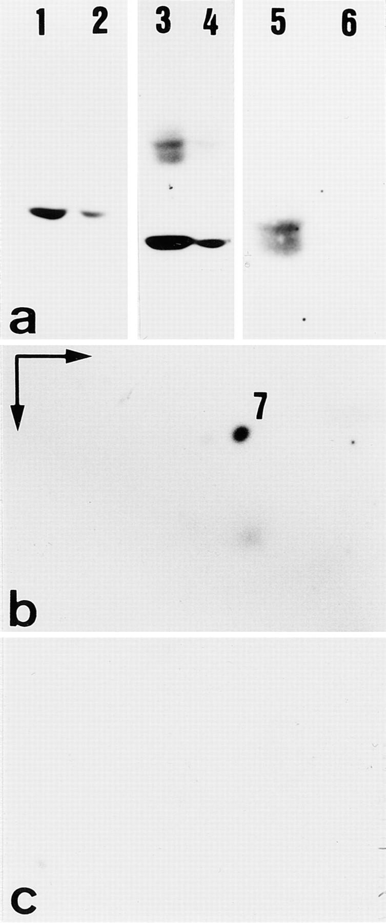
Figure 9.

Altered keratin expression in uterus of K18 null mice. (a–c) Western blot analysis of total proteins separated by SDS-PAGE. (a) Proteins separated by SDS-PAGE. Note the slight reduction of K8 (lanes 1 and 2, reacted with mAB Troma 1) and 19 (lanes 3 and 4, reacted with mAB Troma 3) in extracts from K18 null mice (lanes 2 and 4) compared to those from wild-type mice (lanes 1 and 3). The nature of the upper bands in lane 3 is unknown. (lanes 5 and 6) Detection of K18 with mAB Ks 18.04 in wild-type (lane 5), but not in K18 null animals (lane 6). The doublet in lane 5 probably indicates some degradation. (b and c) Analysis of cytoskeletal proteins from uterus of wild-type (b) and homozygous K18 mice (c) by two-dimensional gel electrophoresis, followed by Western blot analysis. Proteins were separated by isoelectric focusing (IEF) in the first and by SDS-PAGE in the second dimension. Note the presence of K7 in wild-type (b) and its absence in K18 null mice (c) as judged by Western blotting with mAB OVTL 12–30 and enhanced chemiluminescence.
