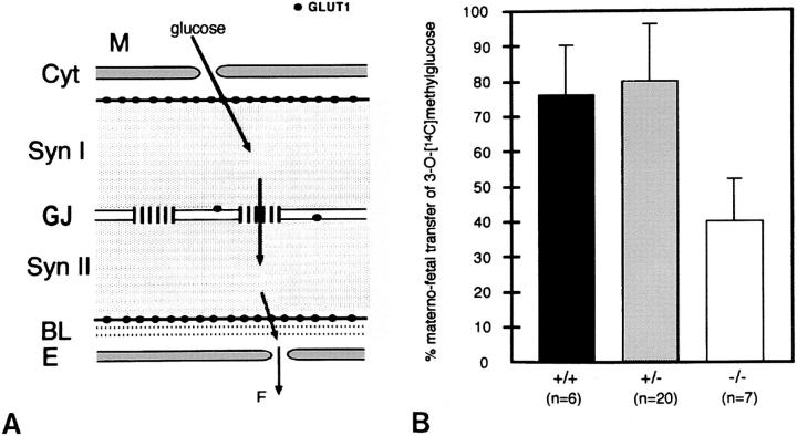
Figure 9.
(A) Schematic drawing of the trophoblast layers of the labyrinth in the murine placenta. The localization of gap junctional Cx26 channels and glucose transporter GLUT1 proteins as well as the likely route of glucose transfer from maternal to fetal blood vessels are illustrated. Glucose enters via GLUT1 the cytoplasm of the syncytiotrophoblast I, diffuses mainly via gap junction channels into syncytiotrophoblast layer II, and is released via GLUT1 at the basal side of the trophoblast membrane into fetal blood circulation through fenestrated endothelial cells (adapted from Takata et al., 1994). M, maternal blood; Cyt, cytotrophoblast; Syn I, syncytiotrophoblastic layer I; GJ, Cx26-containing gap junctions; Syn II, syncytiotrophoblastic layer II; BL, basal lamina; E, endothelial cell; F, fetal blood circulation. (B) Uptake of 3-O-[14C]methylglucose into whole embryos at day 10 pc from maternal blood across the placenta. Significantly decreased accumulation (P < 0.01) of radioactivity was measured in homozygous Cx26-defective (−/−) embryos compared with heterozygous (+/−) and wild-type embryos (+/+). n designates the number of embryos investigated.

