Figure 1.

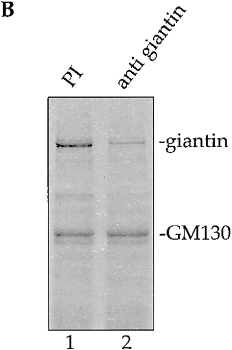
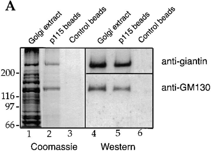
p115 binds to giantin. (A) Rat liver Golgi membranes were solubilized with buffer containing 0.5% Triton X-100, the extract cleared by centrifugation, and then incubated with biotin-p115 immobilized on streptavidin beads, or control beads. The clarified Golgi extract (lanes 1 and 4; 20% of total) and proteins bound to p115 beads (lanes 2 and 5; 50% of total) or control beads (lanes 3 and 6; 50% of total) were fractionated by SDS-PAGE and either stained with Coomassie blue or transferred to nitrocellulose and probed for GM130 and giantin, using specific antibodies. Molecular weight markers are shown in kD. (B) The clarified Triton X-100 extract of Golgi membranes was either preincubated with preimmune serum (PI, lane 1) or with anti- giantin antibodies (lane 2) before incubation with p115 beads. Bound proteins were fractionated by SDS-PAGE and visualized by Coomassie staining. (C) Golgi membranes were incubated for 10 min at 37°C with interphase HeLa cytosol (lane 1), mitotic HeLa cytosol (lane 2), or mitotic HeLa cytosol inactivated by pretreatment with staurosporine (lane 3). After solubilization of the reisolated membranes with Triton X-100, the cleared supernatants were incubated with p115 beads. Bound proteins were fractionated by SDS-PAGE, transferred to nitrocellulose, and then probed for GM130 and giantin.


