
Figure 3.
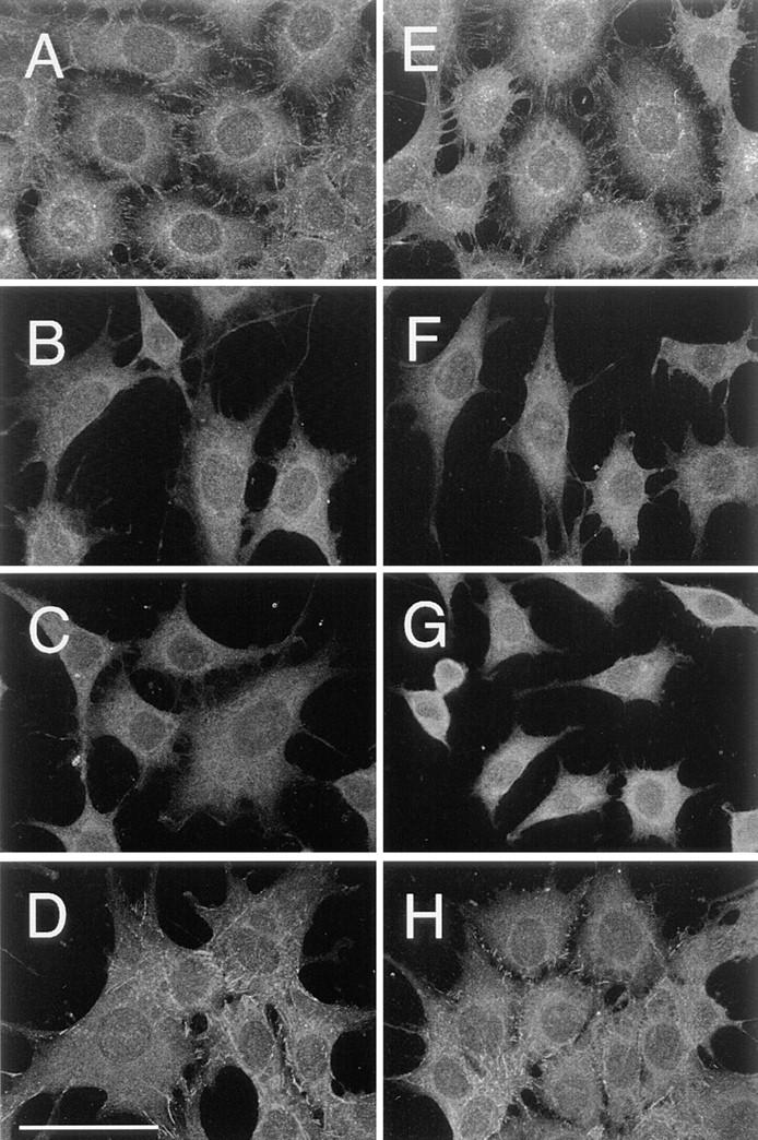
Indirect immunofluorescence staining of E-cadherin on the surface of cells transfected with E-cadherin cDNAs. Cells before (A–D) and after (B–D) incubation with ECCD-2–G40 were fixed with cold methanol, and E-cadherin on the cell surface was stained with ECCD-2 and FITC-labeled anti–rat IgG. Images were obtained by a confocal laser scanning microscopy, with a focal plane at about the height of the dorsal free cell surface near the peripheries of the cell. Staining on the plasma membrane over the nucleus was not observed because it is out of focus. A and E, Wild; B and F, Catenin-minus; C and G, Short-tailed; and D and H, Fusion. Bar, 50 μm.