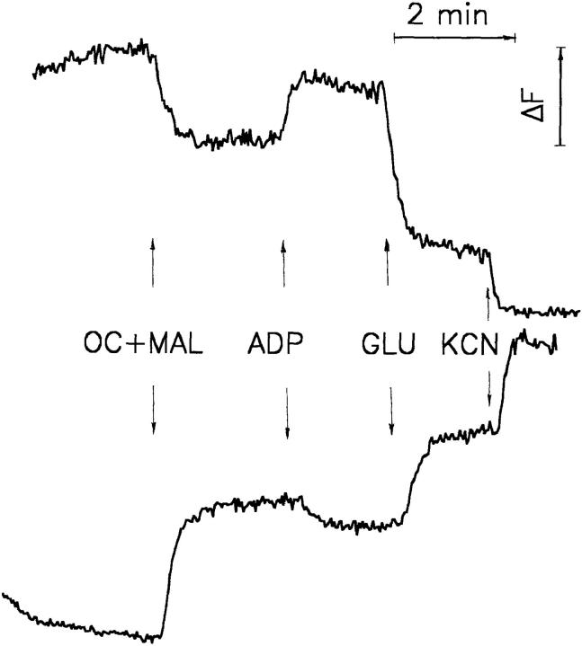
Figure 1.
Plot of the fluorescence changes of NAD(P)H and flavoproteins in bundles of saponin-permeabilized mouse quadriceps muscle fibers. Top trace: flavoprotein fluorescence obtained by 488-nm argon ion laser excitation and registration of the 525-nm emission signal. Bottom trace: NAD(P)H fluorescence obtained by 325-nm HeCd laser excitation and registration of the 450-nm emission signal. Approximately 5 mg of wet weight muscle fibers were attached to glass wool and perfused as described (Kunz et al., 1994). Additions: OC (octanoylcarnitine), 1 mM; MAL (malate), 5 mM; ADP, 1 mM; GLU (glutamate), 10 mM; KCN, 4 mM.

