
Figure 10.
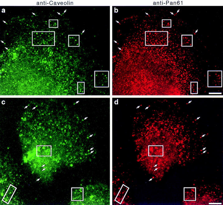
Dynamin localizes to caveolae in cultured hepatocytes. Fluorescence micrographs representing laser scanning confocal microscopy of cultured hepatocytes that were double labeled with a monoclonal anti-caveolin antibody (a and c) as a marker for caveolae and the polyclonal anti-Pan61 antibody (b and d) to label the endogenous dynamin. Two separate fields of cells are shown (top and bottom). A significant number of vesicular structures are labeled with both antibodies (arrows and outlined areas), indicating a colocalization of dynamin and caveolin. A similar staining pattern was obtained with the anti-Pan65 antibody but not in controls where primary antibodies were omitted (not shown). Bars, 8.0 μm.