
Figure 4.
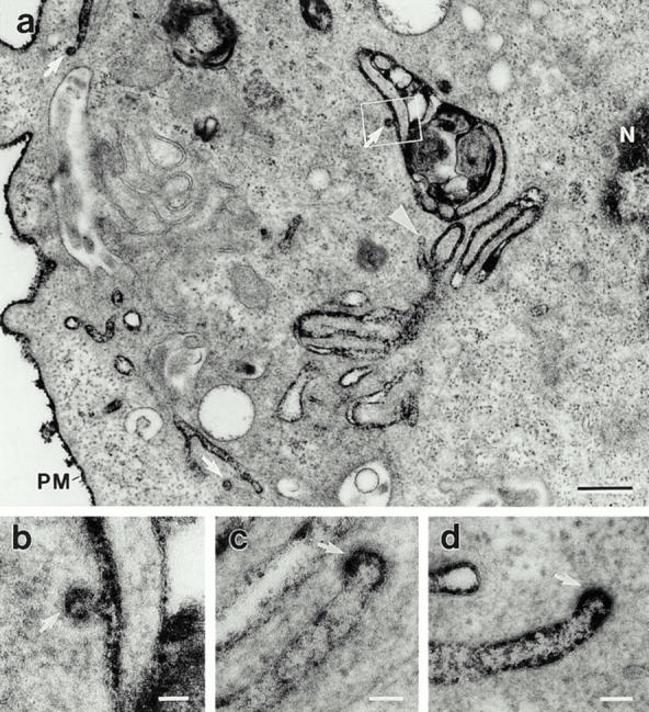
Microinjected anti-dynamin antibodies induce the accumulation of long clathrin-coated pits. Electron micrographs showing the long plasmalemmal invaginations and associated coated pits (arrows) that form in anti-dynamin antibody-injected cells. Hepatocytes were injected with the anti-Dyn2T antibody as in Fig. 2 and then processed for electron microscopy with ruthenium red included in the fixatives to stain the surface membranes. (a) The densely stained structures, although deep within the cytoplasm near the nucleus (N), are continuous with the plasma membrane (PM). A nonclathrin-coated bud that is connected to a plasmalemmal invagination also can be seen (arrowhead). The outlined area is shown at higher magnification in b. (b–d) The bristle-like clathrin coat can readily be seen on the distended regions, or buds, of long plasmalemmal invaginations. Similar invaginations were found when the anti-Pan65 antibody was injected, but not in cells that were injected with the control solutions (not shown). Bars: (a) 0.3 μm; (b–d) 0.05 μm.