
Figure 5.
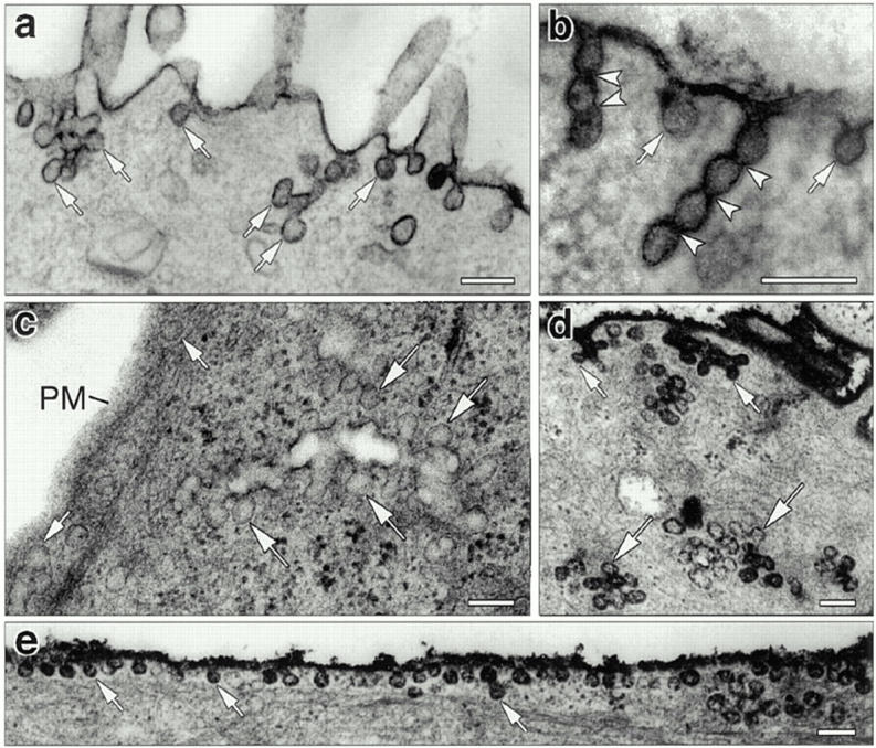
Accumulation of distinct endocytic structures in cultured hepatocytes injected with anti-dynamin antibodies. (a–c) Electron micrographs show typical membrane invaginations found at the surface of cells that were injected with the Dyn2-specific anti-Dyn2T antibody. Numerous budding profiles (small arrows) lacking clathrin coats and resembling caveolae were found at the plasma membrane (a and b). Long “chains” of vesicles separated by constrictions (arrowheads) also were found attached to the plasma membrane (b). In addition, clusters of multiple vesicular profiles were observed deeper within the cell (c, large arrows). Similar structures were found in the anti-Pan65 antibody-injected cells but not in the control-injected cells. (d and e) Electron micrographs of anti-Dyn2T (d) and anti-Pan65 (e) antibody-injected cells that were fixed and stained with ruthenium red as in Fig. 4. Dark vesicles reveal both surface (small arrows) and deep (large arrows) membrane invaginations that are continuous with the plasma membrane. Bars, 0.15 μm.