
Figure 8.
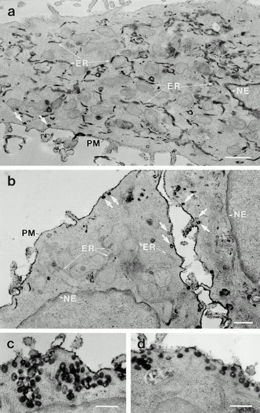
Anti-dynamin antibodies prevent the caveola-mediated endocytosis of HRP–cholera toxin B, as confirmed by electron microscopy. Electron micrographs show hepatocytes that were injected with either a heat-inactivated antibody as a control (a) or the anti-Pan65 antibody (b–d). After the injections, cells were incubated with HRP–cholera toxin B as in Fig. 6, fixed, then processed for diaminobenzidine cytochemistry and electron microscopy. (a) A control-injected cell showing the internal electron-dense peroxidase reaction product that is sequestered largely within elements of the rough endoplasmic reticulum (ER) and the nuclear envelope (NE), with little remaining at the plasma membrane (PM). (b–d) Cells injected with the native anti-Pan65 antibody have little, if any HRP–cholera toxin B labeling of the endosomes, the endoplasmic reticulum, and the nuclear envelope, with most of the peroxidase reaction product residing on the plasma membrane or in numerous caveolae (arrows) and grape-like caveolar clusters. Similar observations were made in cells injected with the anti-Dyn2T antibody (not shown). Bars: (a) 0.5 μm; (b–d) 0.2 μm.