
Figure 10.
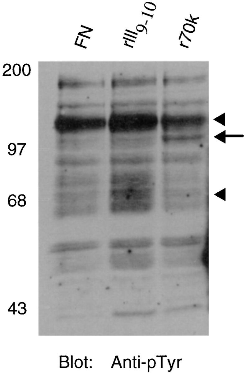
Tyrosine phosphorylation of proteins isolated from cells adherent to r70 kD, rIII9,10, or fibronectin. Cycloheximide-pretreated fibroblasts were resuspended in DME/0.4% ReduSerII containing 20 μg/ml cycloheximide and seeded at 106 cell/ml onto 100-mm tissue culture dishes precoated with fibronectin (FN), rIII9,10, (rIII9 ,10), or r70 kD (r70k). After a 15-h incubation at 37°C, cells were lysed with RIPA buffer, and equal aliquots of clarified lysates were electrophoresed into 8% SDS-PAGE gels. Gels were transferred to nitrocellulose and immunoblotted (Blot) using an anti-phosphotyrosine (Anti-pTyr) antibody. The positions of p125 and p70 are indicated by the arrowheads. The position of p110 is indicated by the arrow to the right of the blot. Molecular mass standards are the same as those indicated in the legend to Fig. 4.