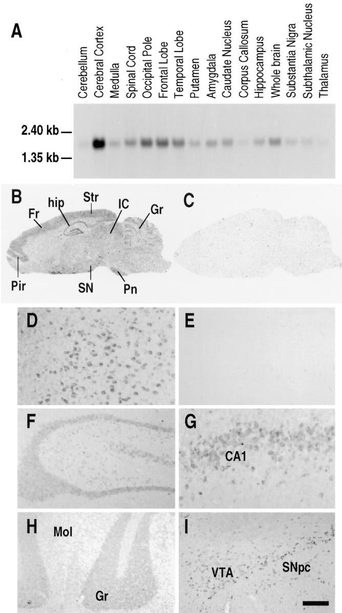
Figure 5.

Expression of Rnd1 within human and rat brain. (A) Northern blot analysis of Rnd1 expression in different regions of the human brain. (B–C) Photomicrographs of the autoradiograms of sagittal rat brain sections after in situ hybridization histochemistry with (B) the antisense Rnd1 riboprobe and (C) the corresponding sense riboprobe. Exposure time is 21 d. (D–I) High power bright-field microphotographs of coronal rat brain sections immunostained with the (D and F–I) anti-Rnd1 antibodies, and (E) anti-Rnd1 antibodies after preincubation with the antigenic peptide. (D and E) cerebral cortex, (F) hippocampus, (G) CA1 pyramidal neurons, (H) granular cell layer of the cerebellar cortex, and (I) VTA and substantia nigra pars compacta. Cx, cerebral cortex; Fr, frontal cortex; Gr, cerebellar granular layer; hip, hippocampus; IC, inferior colliculus; Pir, piriform cortex; Pn, pontine nuclei; SN, substantia nigra; and Str, striate cortex. Bar: (D, E, and G) 65 μm; (F, H, and I) 200 μm.
