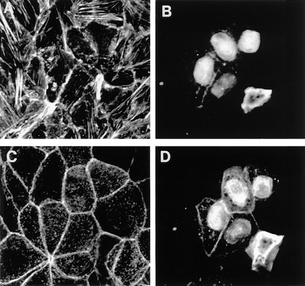
Figure 9.
Effects of Rnd1 expression on actin filaments in epithelial cells. Confocal analysis of MDCK cells microinjected with a pRK5 vector expressing wild-type Rnd1. Cells were prepermeabilized, fixed 3 h after injection, and then stained to show Rnd1 expression (B and D) and actin filaments (A and C). Images A and B show a confocal section through a region juxtaposed to the basal membrane, and images C and D represent a section through the adherens junction lateral membranes. We are unable to assess whether there is any significance to the nuclear staining pattern of overexpressed Rnd1. It has often been observed that overexpression of other small G proteins leads to nuclear localization of the posttranslationally unprocessed protein.

