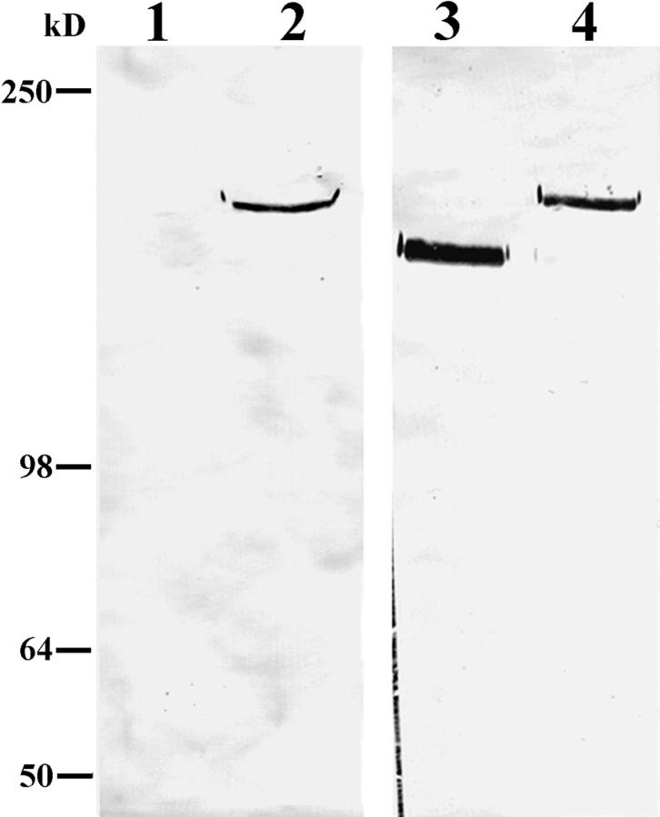
Figure 3.
Approximately 10 μg of either MCF-10A (lanes 1 and 3) or pp126 cell (lanes 2 and 4) ECM was run on lanes of a 6% gel, transferred to nitrocellulose, and then processed for immunoblotting using either a mouse serum (Cta3) raised against residues 1561–1713 at the COOH terminus of the α3 subunit of laminin-5 (lanes 1 and 2) or the α3 subunit monoclonal antibody 10B5 (lanes 3 and 4). Antiserum Cta3 recognizes a 190-kD protein in pp126 ECM (lane 2), but does not show reactivity with any polypeptide in MCF-10A ECM (lane 1). The 190-kD species in pp126 ECM is also recognized by 10B5 antibodies (lane 4). However, unlike the Cta3 serum, 10B5 antibodies recognize a 160-kD protein in MCF-10A ECM (lane 3). Molecular weight markers are indicated at the left of the immunoblots.

