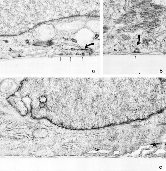
Figure 7.

SCC12 cells were maintained for 24 h on plasmin-modified (1 μg/ml for 90 min) pp126 matrix (a), plasmin-modified purified pp126 laminin-5 (b), and plasmin-modified pp126- derived laminin-5 that had been incubated for 30 min at 37°C with 50 μg/ml of the laminin-5 function-inhibiting antibody 1947 before addition of the cells (c). The SCC12 cells were then processed for electron microscopy and cross-sections of the cells were prepared. In a and b, several hemidesmosomes in the SCC12 cells are observed at sites of cell– matrix association (arrows). Each has a triangular, trilayered plaque that is associated with intermediate filaments (curved arrows in a and b). c, the SCC12 cell has assembled no obvious hemidesmosomes along regions of cell–matrix interaction. The arrow indicates one hemidesmosome-like structure that is not associated with the substrate. This structure may even be a half-desmosome. a and c are at the same magnification. Together, these results provide evidence that plasmin-modified pp126 cell laminin-5 is capable of inducing the assembly of hemidesmosomes. Bars: (a and c) 500 nm; (b) 200 nm.
