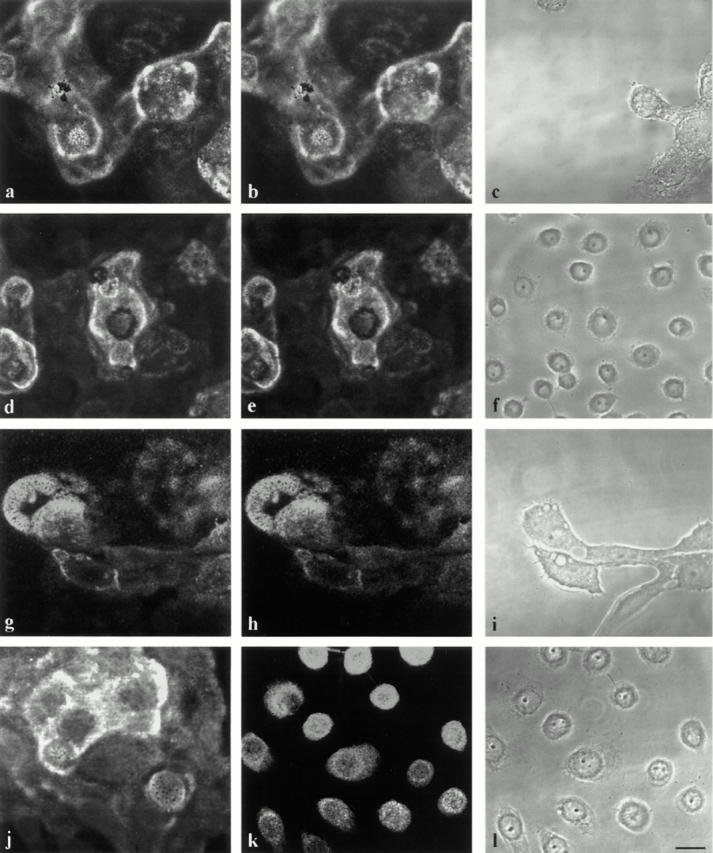
Figure 8.

MCF-10A cells (a–c and g–i) and pp126 cells (d–f and j–l) were processed for indirect double-label immunofluorescence microscopy using antibodies against laminin-5 (a, d, g, and j) in combination with either an antibody against plasminogen (b and e) or tPA (h and k). Note that there is colocalization of plasminogen and laminin-5 staining patterns in a and b as well as d and e. In g and h, tPA shows codistribution with laminin-5. In k, tPA localizes to cell bodies but does not colocalize with laminin-5 in j. c, f, i, and l are phase images. Bar, 25 μm.
