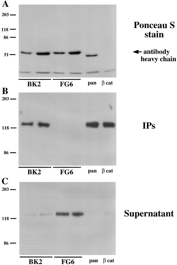
Figure 1.
Coimmunoprecipitation of PTPμ and cadherin from lysates of MvLu cells. Lysates of MvLu cells were subjected to immunoprecipitation with two different concentrations of anti-PTPμ antibody BK2 or an isotype-matched antibody to PTP1B (FG6), as well as the pan-cadherin and anti–β-catenin antibody. The relative amounts of antibody heavy chain in each immunoprecipitate are shown in a Ponceau S stain of the immunoblot (A). Immunoblots using pan-cadherin antibody were performed on the immunoprecipitates (B). The quantity of cadherin remaining in the supernatant after immunoprecipitation was assessed by immunoblotting (C). The results illustrate that the majority of cadherin in the lysate coimmunoprecipitated with PTPμ.

