
Figure 1.
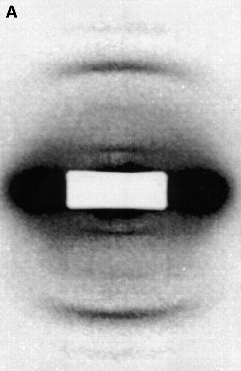

(A) X-ray diffraction image of fibrillin-rich zonular filaments in the presence of Ca2+. Data were recorded on beamline 2.1 CLRC Daresbury (camera length, 6.25 m); experiments were conducted at 20°C. The predominant first and third orders can be seen. The second and fourth diffraction intensities are very weak. The diffraction image has been corrected for the detector response and a suitable empty cell image removed. Intensities were scaled within individual minimum–maximum boundaries. (B) X-ray diffraction image of fibrillin-rich zonular filaments in the absence of Ca2+. Ca2+ ions have been removed by immersion in a buffer containing an excess of EGTA. In this case, the relative intensities of the diffraction peaks can be seen to decrease as a function of diffraction order. However, the absolute intensity of the third order was found to remain relatively constant relative to Ca2+-containing samples, and the major effects are the enhancement of intensity for the first and second orders with EGTA added.