Figure 4.
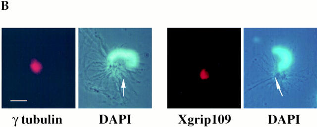
Xgrip109 is localized to centrosomes. (A) Xgrip109 and γ tubulin colocalized to the centrosomes in XLK-WG cells. γ tubulin, γ-tubulin localization revealed by fluorescein secondary antibody; Xgrip109, Xgrip109 localization revealed by Cy3 secondary antibody; DAPI, DNA staining with DAPI; Nom, Nomarski images of the cells. (B) Xgrip109 is localized to the in vitro–assembled centrosomes. The in vitro–assembled centrosomes were spun onto glass coverslips and indirect immunofluorescence staining was carried out using anti–γ-tubulin and anti-Xgrip109 antibodies (refer to Materials and Methods). γ tubulin, γ tubulin was localized to the tip of the sperm nucleus; Xgrip109, Xgrip109 was also localized to the tip of the sperm nucleus; DAPI, the two-sperm nuclei that were stained with either anti–γ-tubulin antibody (GTU-88) or anti-Xgrip109 antibodies (109-2) were stained with DAPI. The images are a combination of fluorescence and phase images. Arrows, microtubule asters at the tips of the two-sperm nuclei. Bars: (A) 20 μm; (B) 10 μm.


