
Figure 2.
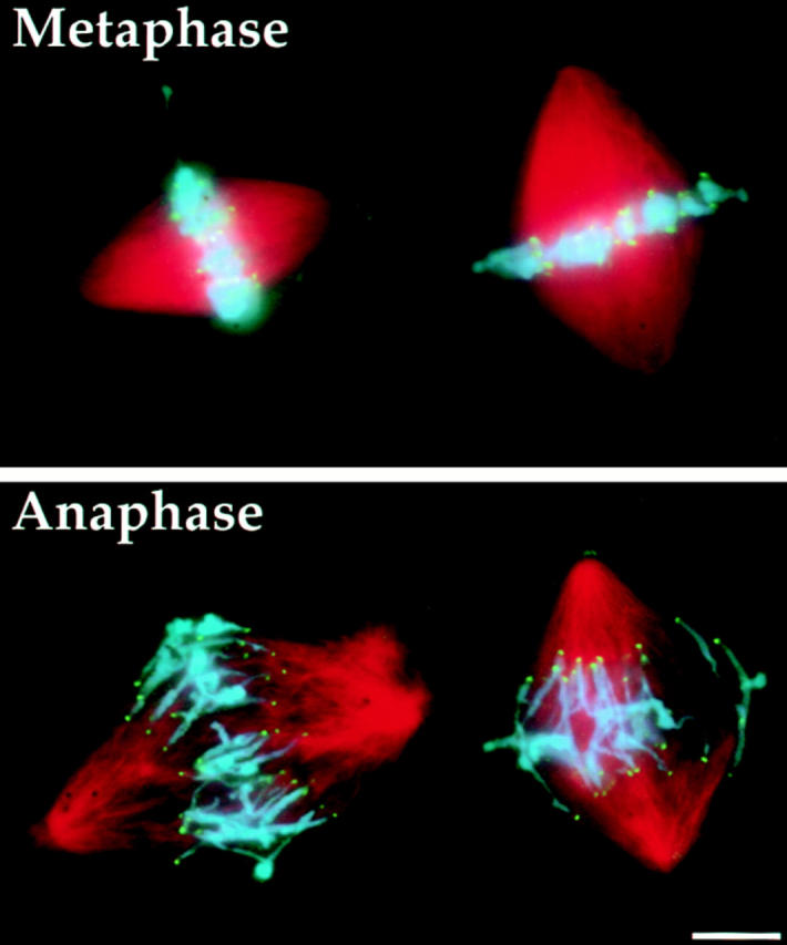
Immunofluorescence micrographs of fixed spindles showing that kinetochores lead chromosomes-to-pole movement during anaphase in Xenopus extract spindles. Spindle MTs are stained red using rhodamine tubulin, chromosomes are stained blue by DAPI and CENP-E is stained green using an anti– CENP-E primary and fluorescein-labeled secondary antibodies. Metaphase-arrested bipolar spindles with replicated chromosomes contained tight metaphase plates with sister kinetochores of bivalent chromosomes localized to the equatorial region of the spindle (top). Addition of a pulse of calcium to the extract inactivated the metaphase arrest and 8 min after calcium addition separated sister chromatids were seen moving poleward (bottom). Discrete foci of CENP-E are clearly visible at the leading edges of the poleward-migrating chromosomes. Bar, 10 μm.