
Figure 3.
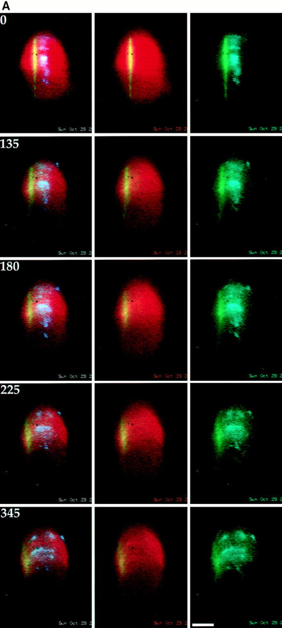

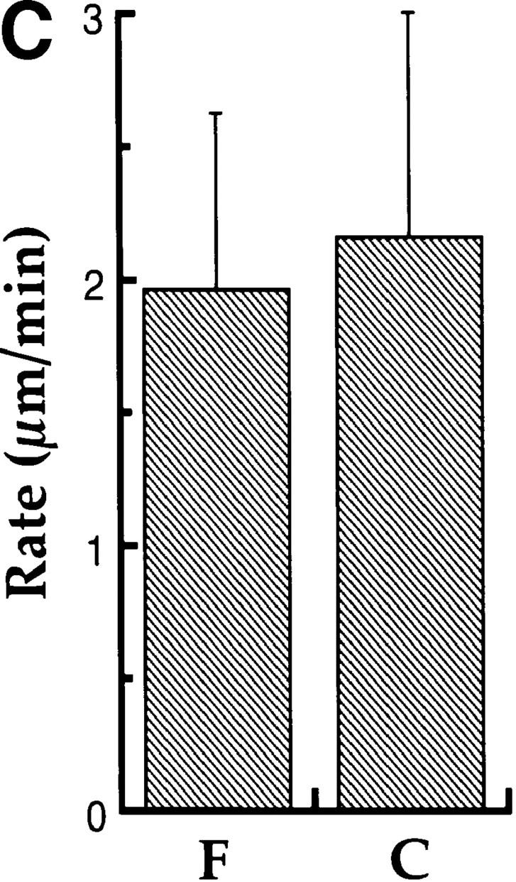
Simultaneous observation of chromosome movement and poleward MT flux in Xenopus extract spindles. (A) Panels from a sequence showing the similarity of poleward MT flux and chromosome movement during anaphase in Xenopus extract spindles. X-rhodamine tubulin-labeled spindle MTs are red, the photoactivated C2CF tubulin–containing MTs on the spindle MT lattice are green and the DAPI-labeled chromosomes are blue. The left column shows three-color overlays of the spindle, the mark and the chromosomes; the middle column shows two-color overlays of the fluorescent mark on the spindle MTs; and the right column shows two-color overlays of the chromosomes and the fluorescent mark. Time elapsed after the mark was made is indicated in seconds on the top left corner of the three-color overlay panels. For this particular sequence the mark was made 11 min after addition of calcium to trigger anaphase. (B) Fluorescence intensity linescan analysis of chromosome-to-pole movement and poleward MT flux during anaphase in Xenopus extract spindles. Fluorescence intensity along the spindle axis in the fluorescein channel is plotted for four different time points for the sequence in Fig. 3 A. The position of the leading edge of the chromosomes, obtained from linescans in the DAPI channel, is indicated by dots on the fluorescence intensity profiles of the mark. The left spindle pole is located at the x axis origin. The fluorescent mark, which was made close to the leading edges of the chromosomes, moved poleward and decayed in intensity, presumably as a result of spindle MT turnover. The mark also broadened as it moved, indicating the existence of differentially fluxing spindle MT subpopulations. (C) Summary of the analysis of rates of poleward MT flux (F) and chromosome-to-pole movement (C) during anaphase in Xenopus extract spindles. The plotted values represent the mean ± SD for rates measured on 11 spindles. Bar, 20 μm.