
Figure 4.
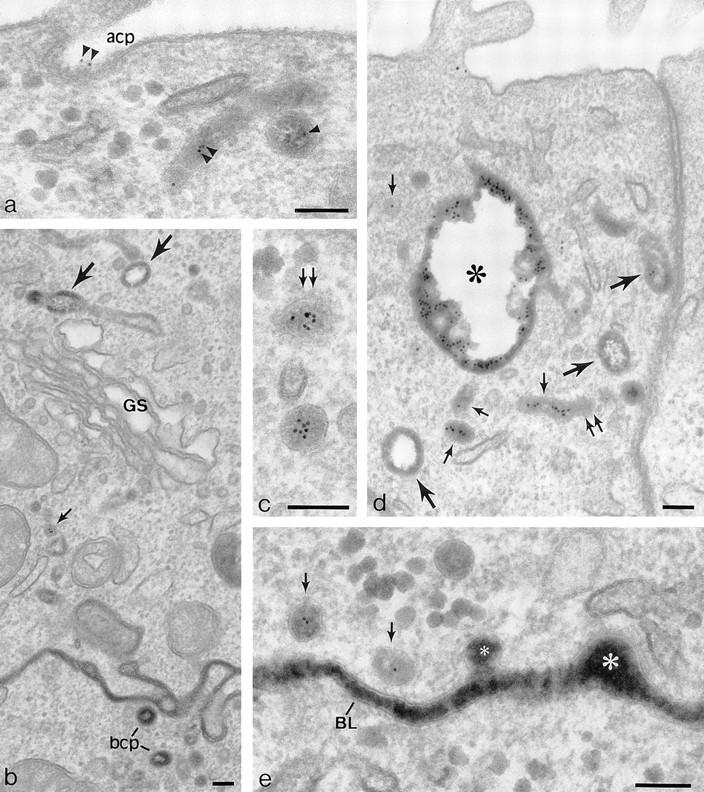
Penetration of gold-labeled TR from the apical surface into transcytotic elements preloaded with TF-HRP from the basolateral surface. (a) At the apical border, apically applied gold-labeled TR are present in coated pits (acp) and have gained access to endosomal elements (small arrowheads) containing TF-HRP that has been loaded from the basolateral surface. (b) At the basolateral border, TF-HRP is distributed in the basolateral space and in coated pits/vesicles (bcp). Deeper in the cell, the TF-HRP is present in tubules and vacuoles of variable diameter (arrows) and in small (60-nm-diam) basolateral vesicles (small arrow) that also contain apically derived gold-labeled TR. (c) Two 60-nm-diam vesicles (one coated, double arrows) showing (1) the homogeneous distribution of DAB reaction product due to TF-HRP derived from the basolateral border and (2) the central cluster of gold-labeled TR (internalized from the apical surface) that characterize the content of basolateral vesicles. (d) Endosomal vacuole (*) surrounded by tubules and vesicles containing apically and basolaterally derived tracer. Large arrows indicate larger tubules with a peripheral distribution of DAB reaction product similar to the vacuole. Small arrows indicate 60-nm-diam tubules and vesicles filled with DAB reaction product. In cross-sections of these narrow tubules, the gold particles occupy a central position, which in longitudinal sections are often seen in single file. Double arrows indicate a clathrin-coated bud. (e) Basolateral border (BL) showing 60-nm-diam basolateral tubules/vesicles (arrows) containing apically derived gold TR tracer and a smooth surfaced invagination (small asterisk) where an exocytotic vesicle has fused. The density of the DAB reaction product within the exocytotic invagination is similar to that given by the TF-HRP in the lateral space (and much stronger than in the unfused vesicles in the peripheral cytoplasm), suggesting that there is a rapid equilibration of intra- and extracellular tracer when vesicles fuse. The large asterisk indicates an endocytotic invagination identifiable because it is larger (∼100-nm-diam) and is clathrin-coated. Bars, 0.1 μm.