
Figure 6.
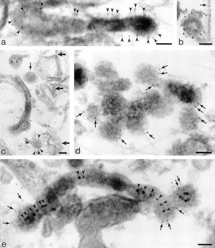
Labeling cytoplasmic domains of TR and coat proteins on 60-nm-diam tubules and vesicles loaded basolaterally with TF-HRP in polarized monolayers permeabilized with digitonin. (a) Thick section showing distribution of H68.4-labeled TR in a 100-nm-diam form of tubule that extends into a 60-nm-diam form with a terminal bud. Permeabilized cell treated with Tris and Triton X-100 to remove clathrin lattices. TF-HRP reaction product and the gold labeling (arrowheads) indicates some concentration as the tubule narrows. (b) 60-nm-diam vesicle containing internalized TF-HRP. The cytoplasmic domains of the TR in this free vesicle are labeled with 5-nm H68.4 antibody. Arrow indicates the plasma membrane. (c) Thin section showing distribution of H68.4–gold on TF-HRP–containing endosomal elements (asterisks) and basolateral vesicles. One basolateral vesicle is coated and unlabeled with gold (small arrow) and the other is labeled (arrowheads). Large arrows indicate basolateral plasma membrane. (d) Thick section showing 60-nm-diam endosomal tubules labeled for clathrin (small arrows). (e) Thick section of 60-nm-diam endosomal tubules labeled for γ-adaptin with 5-nm-gold (small arrows). The tubules contain apically derived TR gold tracer in linear array (arrowheads) and the coated bud contains a central cluster of six particles. Bars, 0.05 μm.