
Figure 8.
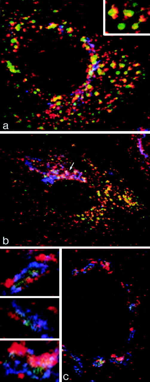
Immunofluorescent localization of γ-adaptin in cells grown on solid substrata. (a and b) MDCK cells stably expressing ST-HRP were transiently transfected with human TR and were incubated for 1 h at 37°C with TF-FITC. They were triple labeled for γ-adaptin (red), TF-FITC (green), and ST-HRP (blue). (a) In the peripheral cytoplasm, γ-adaptin is closely associated with endocytosed TF-FITC. In the enlarged structures shown in the inset, γ-adaptin appears to cap the tubules extending from the TF-FITC–containing vacuoles. (b) In the Golgi region, the γ-adaptin is closely associated with ST-HRP (arrow), but overlap between TF-FITC and the ST-HRP is negligible. (c) Triple label for γ-adaptin (red), ST-HRP (green), and β′-COP (blue). In addition to labeling the punctate structures in the peripheral cytoplasm, γ-adaptin is closely associated with β′-COP label in the ST-HRP– containing Golgi elements. Seen at high magnification (insets), it is clear that the Golgi elements contain discrete domains in which either γ-adaptin or COP1 are concentrated.