
Figure 8.
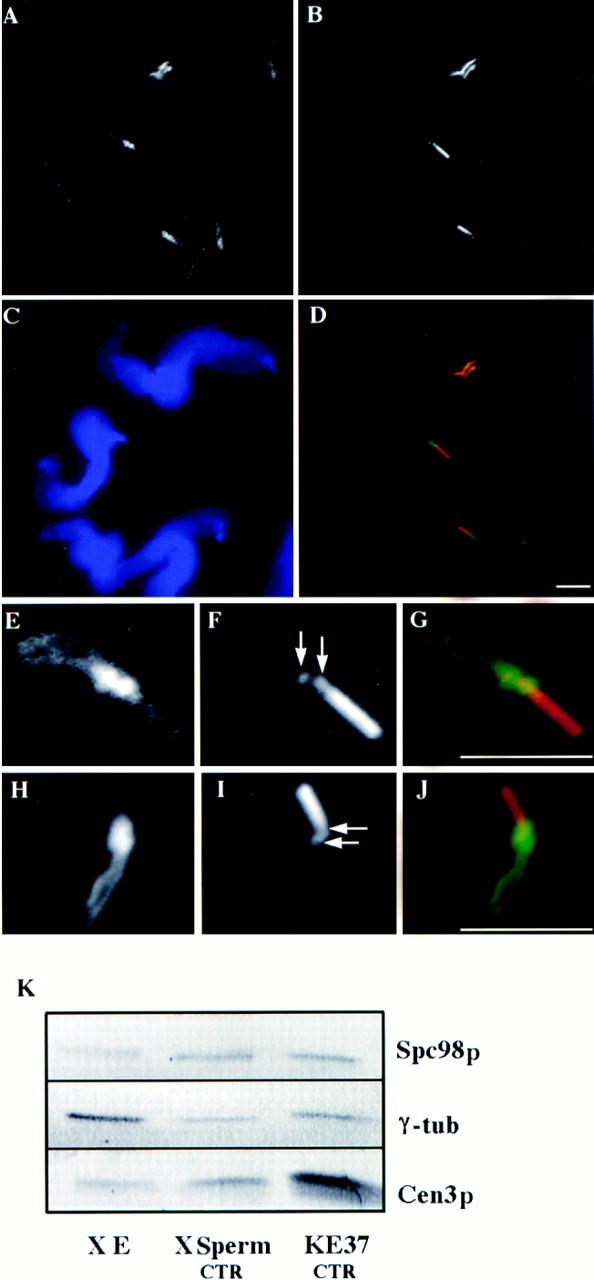
(A–J) Decoration of Xenopus sperm centrosome before activation in Xenopus egg extract. Double labeling with either anti–γ-tubulin antibodies (A and E) or with anti-HsSpc98p (H) and with GT335, a monoclonal antibody recognizing polyglutamylated tubulin (B, F, and I; arrows point to the centrioles). D, G, and J are the superposition of the respective labelings. (C) DAPI staining of the sperm nuclei. (A–D) A field of Xenopus sperm heads. (E–J) High magnification of a single sperm head. Note that both γ-tubulin and Spc98p accumulate around the two centrioles and along the striated rootlets. (K) Western blot analysis of Xenopus egg extract (X E), Xenopus sperm centrosomes (X Sperm CTR), and human somatic centrosomes (KE37 CTR). Proteins were probed with affinity-purified HsSpc98p IgG, anti–γ-tubulin antibody, or anti-HsCen3p antibody. Note the presence of similar amounts of both Spc98p and γ-tubulin in Xenopus sperm centrosomes and human somatic centrosomes. Bars, 5 μm.