
Figure 9.
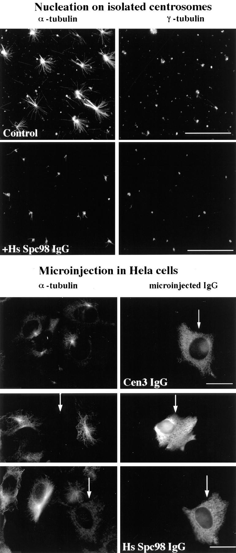
Affinity-purified anti-HsSpc98p antibodies inhibit microtubule nucleation in vitro (top) and in vivo (bottom). (Top) Microtubule nucleating activity of isolated centrosomes was monitored by double immunofluorescence with anti–α-tubulin and with anti–γ-tubulin. Centrosomes were preincubated with preimmune immunoglobulins (Control) or with anti-HsSpc98p immunoglobulins (+HsSpc98 IgG) on ice for 30 min and subsequently incubated in PC-tubulin. Microtubules were allowed to grow for 4 min. Note the specific inhibition of aster formation after 4 min of growth with HsSpc98 immunoglobulins, while the control centrosomes were growing typical microtubule asters. (Bottom) Anti-HsSpc98 immunoglobulins were microinjected into HeLa cells (HsSpc98 IgG). 2 h after microinjection, microtubules were depolymerized for 2 h with 5 × 10⁻⁶ M nocodazole. After washing out the nocodazole, microtubules were allowed to regrow for 10 min. Cells were fixed with methanol and processed for immunofluorescence with monoclonal anti–α-tubulin antibodies followed by the mouse and rabbit secondary antibodies. In this way, only the microinjected cells are detected with the fluorescein anti–rabbit secondary antibody. Note that the nonmicroinjected cells presented the usual centrosome-growing microtubule aster, while heavily microinjected cells did not show any microtubule (arrows). Note that in cells micronjected with an unrelated antibody (anti-Cen3p IgG), microtubule regrowth is not affected. Bars, 10 μm.