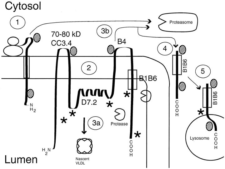
Figure 11.
Model of apoB structure in the membrane of the endoplasmic reticulum. The diagram shows a putative model of nascent apoB in the ER membrane. Pathways are explained in the text. Putative epitopes for the polyclonal anti-LDL antibodies are shown (★). The shaded ovals represent chaperon proteins, including heat shock protein 70. The circles with a missing wedge represent putative proteases in the ER.

