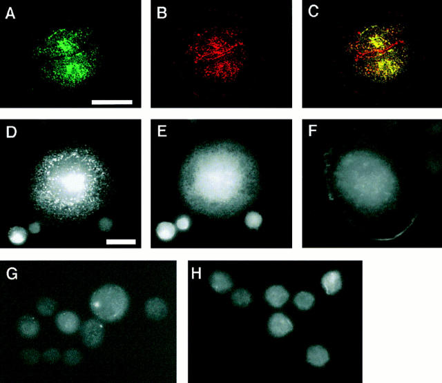
Figure 3.
Clustering activity of wild-type and mutant C-cadherin molecules. CHO cells stably expressing C-cadherin or mutant molecules were allowed to attach for 1 h to glass substrata coated with CEC1-5 (A–E, G, and H) or poly-l-lysine (F) and then fixed and stained for the cadherin molecule (A, C, D, F, G, and H) or β-catenin (B, C, and E) by simultaneous dual-label immunofluorescence microscopy. Wild-type C-cadherin was detected using a polyclonal antibody raised against the whole cytoplasmic tail of mouse E-cadherin; mutants CT, CT669, and CT-CAT were detected by staining for the myc epitope tag. Specimens A–C were examined by confocal laser scanning microscopy with the plane of focus at the cell– substrate interface; all other samples were examined by epi-illumination microscopy. (A–C) Wild-type C-cadherin localized in clusters (A) that colocalized with β-catenin (B), as seen by the yellow fluorescence in the overlay image (C). Some β-catenin that did not colocalize with C-cadherin was detected at borders where the cells were close but not directly touching; this is likely to be in a cytoplasmic pool. (D and E) CT669 clustered in cells attached to substrata coated with CEC1-5 (D), but not with poly-l-lysine (F). CT669 clusters did not colocalize with β-catenin that remained diffusely distributed within cells (E). The image in E was overexposed for clarity in reproduction and therefore may give a misleading impression of β-catenin expression in CT669-CHO cells, which was similar to untransfected CHO cells (Fig. 2). Neither CT (G) nor CT-CAT (H) clustered upon attachment to CEC1-5–coated substrata. Note that CT-CHO and CT-CAT-CHO cells spread less on CEC1-5 than either C-CHO or CT669-CHO cells. Bars: (A–C) 25 μm; (D–H) 10 μm.

