
Figure 11.
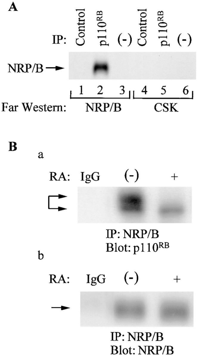
In vitro and in vivo association of NRP/B with p110RB. (A) Lysates of SH-SY5Y cells were immunoprecipitated with anti-p110RB antibody (lanes 2 and 5), analyzed by 7.5% SDS-PAGE, and transferred to Immobilon-P membranes. The membranes were processed for Far Western blotting with the Csk kinase (as a negative control) and NRP/B-purified proteins. Lanes 1 and 4, immunoprecipitations were performed with control antibody; lanes 3 and 6, control Sepharose beads alone. (B) In vivo association of NRP/B with p110RB: (a) 500 μg of total cell lysates obtained from retinoic acid (10 μM) treated and untreated SH-SY5Y cells were subjected to immunoprecipitation with NRP/B and blotted with monoclonal p110RB antibody. Two different migrated forms were observed: The slower migrated form (lane 1) represents the hyperphosphorylated p110RB, and the faster migrated form (lane 2) represents the hypophosphorylated p110RB. (b) After deprobing the blot in a, the membrane was probed with polyclonal NRP/B antibody.