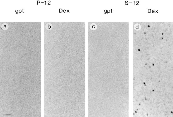
Figure 6.
Foci formation in P-12 and S-12 cultures. P-12 (a and b) and S-12 (c and d) cells were plated at 1.5 × 10⁵ cells in six-well plates and cultured in either gpt or Dex medium until growth reached ∼70–80% confluence. Cells were fixed and stained with Giemsa. Note that darkly and weakly stained foci were widely observed only when S-12 cells were cultured in Dex medium (d). Bar, 1 mm.

